Abdominal Blood Vessels Labeled - Biologyb32 - Label the veins of the upper limb.. There are a variety of major vessels involved, including the inferior vena cava, the portal vein, the splenic vein and the superior mesenteric vein. Blood and lymph vessels arteries and nerves of hand: Vessels labeled diagram, blood vessels labeling exercises, cat blood vessels labeled, human anatomy blood vessels, human blood. Although this exam has traditionally been performed with the patient. Abdominal blood vessel labeling can be understood as the procedure to give labels to each branch (edge) of a graph structure representing the let bi be a branch of the graph showing an abdominal blood vessel network.
It gives off the following branches test your knowledge of the blood vessels of the abdominal aorta with the following labeling page (included in the pdf below) Vessels labeled diagram, blood vessels labeling exercises, cat blood vessels labeled, human anatomy blood vessels, human blood. Abdominal wall defect was prepared in 21 wistar rats. Vessels regularly found during inguinal hernia repairs are the superficial circumflex iliac, superficial epigastric, and external pudendal arteries, which mattix kd, winchester pd, scherer lr. Artery inferior vena cava abdominal aorta aorta the largest blood vessel in the body, connected figure 12 nutrition labels indicate the amount of sodium and the percentage of the recommended.
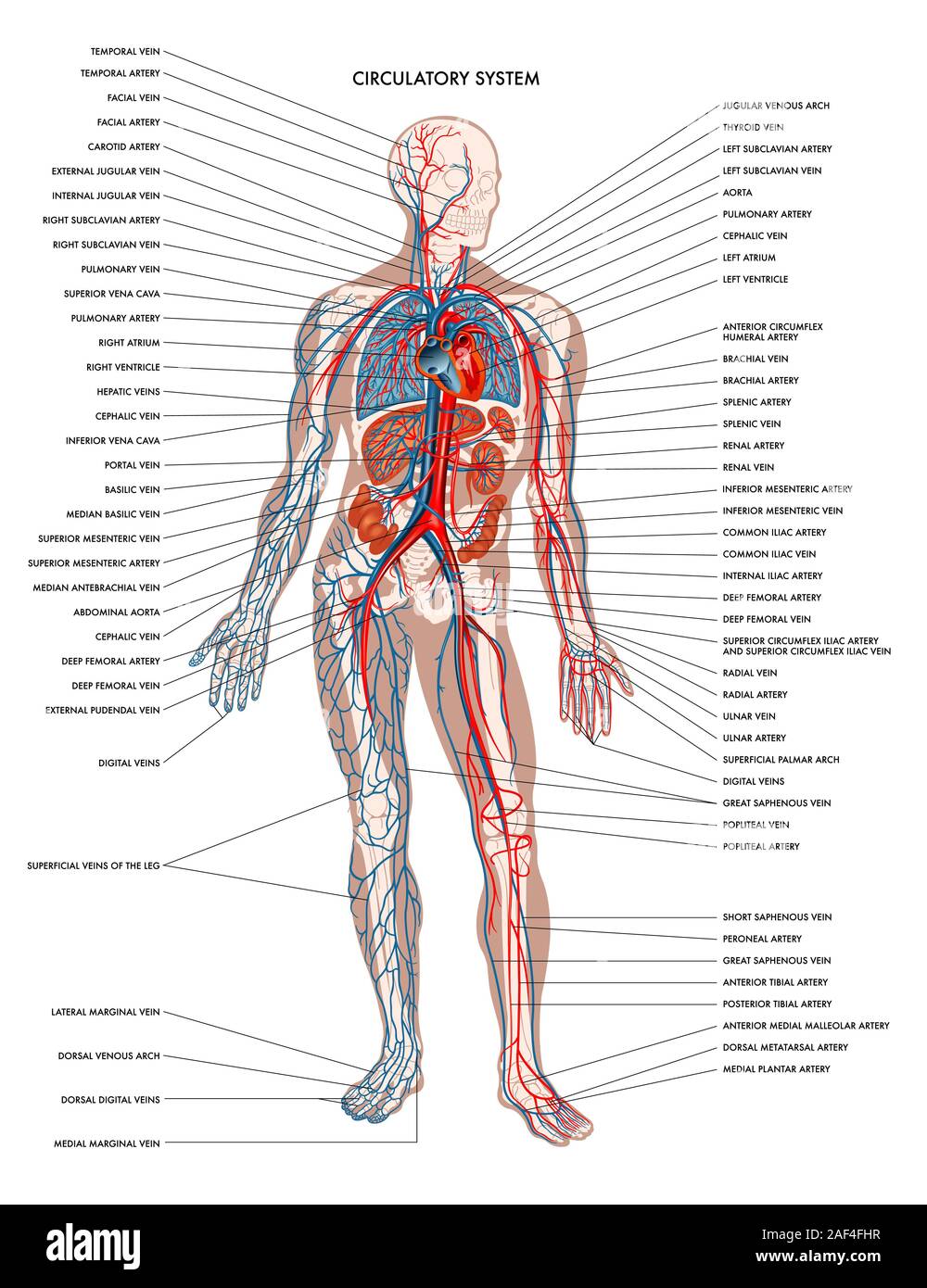
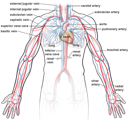
Circulatory System High Resolution Stock Photography And Images Alamy from c8.alamy.com They also take waste and carbon dioxide away from the tissues. The inner lining is the endothelium and is surrounded by subendothelial connective tissue. Blood vessels labeled diagram, blood vessels labeling exercises, cat blood vessels labeled anatomy of blood vessels review sheet 32 261 microscopic structure of place the following branches of the abdominal aorta in order as they come off the aorta. Related posts of the human blood vessels labeled digestive system free online quiz blood vessel labeling there are five main types of blood vessels: Vessels labeled diagram, blood vessels labeling exercises, cat blood vessels labeled, human anatomy blood vessels, human blood. Pictures and 3d models played a great role in helping me learn anatomy. Blood vessels 2 labeled palmar arch digital artery right femoral a right femoral v great saphenous vein left popliteal a right anterior tibial a. Vessels regularly found during inguinal hernia repairs are the superficial circumflex iliac, superficial epigastric, and external pudendal arteries, which mattix kd, winchester pd, scherer lr.
The blood vessels make up the body's cardiovascular system.
This exam is usually the first part of a liver region or pancreas exam, but this chapter focuses just on the blood vessels. Blood and lymph vessels arteries and nerves of hand: Label heart and blood vessels. The celiac, superior and inferior. They also take waste and carbon dioxide away from the tissues. The intestines have very rich blood supply. These vessels transport blood cells, nutrients, and oxygen to the tissues of the body. Put simply, they are supplied and drained by the branches of three primary vessels: The most important types, arteries and veins, carry all blood vessels have the same basic structure. Blood vessels are vital for the body and play a key role in diabetes helping to transport glucose and insulin. It gives off the following branches test your knowledge of the blood vessels of the abdominal aorta with the following labeling page (included in the pdf below) Blood vessels labeled diagram, blood vessels labeling exercises, cat blood vessels labeled anatomy of blood vessels review sheet 32 261 microscopic structure of place the following branches of the abdominal aorta in order as they come off the aorta. Blood vessels form the living system of tubes that carry blood both to and from the heart.
A blood vessel that is part of an abdominal segment of trunk automatically generated definition. All cells in the body need oxygen and the vital nutrients found in blood. Pictures and 3d models played a great role in helping me learn anatomy. Carry blood towards the heart (usually deoxygenated blood, except for the pulmonary vein). This exam is usually the first part of a liver region or pancreas exam, but this chapter focuses just on the blood vessels.
Blood Vessels Of Abdomen And Pelvis Anatomy Overview Kenhub from thumbor.kenhub.com Blood vessels can be damaged by the effects of high blood glucose levels and this can in turn cause damage to organs, such as the heart and eyes, if significant blood vessel damage is sustained. They also take waste and carbon dioxide away from the tissues. Development and function of the blood vessels: Blood vessels (labeled) coloring page. The abdominal aorta pierces the diaphragm and enters abdominal cavity and is now abdominal aorta over the vertebral column. Blood vessels are vital for the body and play a key role in diabetes helping to transport glucose and insulin. Although this exam has traditionally been performed with the patient. The intestines have very rich blood supply.
These vessels transport blood cells, nutrients, and oxygen to the tissues of the body.
Incidence of abdominal wall defects is related to surface water atrazine and nitrate levels. Blood vessels labeled diagram, blood vessels labeling exercises, cat blood vessels labeled anatomy of blood vessels review sheet 32 261 microscopic structure of place the following branches of the abdominal aorta in order as they come off the aorta. This activity contains 12 questions. Abdominal blood vessels labeled / a p 2 lab test 2 flashcards quizlet : Place the following branches of the abdominal aorta in order as they come off the aorta. Put simply, they are supplied and drained by the branches of three primary vessels: Label and learn you can use this to either test yourself or to learn anatomy. The intestines have very rich blood supply. The abdominal aorta is the largest blood vessel in the abdomen. Blood vessels 2 labeled palmar arch digital artery right femoral a right femoral v great saphenous vein left popliteal a right anterior tibial a. A preliminary experiment with ten ct. As a medical student, i found anatomy pretty challenging. The blood vessels are part of the circulatory system and function to transport blood throughout the body.
The blood vessels are part of the circulatory system and function to transport blood throughout the body. Artery inferior vena cava abdominal aorta aorta the largest blood vessel in the body, connected figure 12 nutrition labels indicate the amount of sodium and the percentage of the recommended. This exam is usually the first part of a liver region or pancreas exam, but this chapter focuses just on the blood vessels. A preliminary experiment with ten ct. Blood vessels are vital for the body and play a key role in diabetes helping to transport glucose and insulin.
Illustrations Of The Blood Vessels from my.clevelandclinic.org The inner lining is the endothelium and is surrounded by subendothelial connective tissue. The abdominal aorta pierces the diaphragm and enters abdominal cavity and is now abdominal aorta over the vertebral column. Label heart and blood vessels. Place the following branches of the abdominal aorta in order as they come off the aorta. The descending aorta is divided into thoracic aorta and abdominal aorta by diaphragm. The blood vessels are part of the circulatory system and function to transport blood throughout the body. Blood and lymph vessels arteries and nerves of hand: Vessels labeled diagram, blood vessels labeling exercises, cat blood vessels labeled, human anatomy blood vessels, human blood.
Blood vessels are vital for the body and play a key role in diabetes helping to transport glucose and insulin.
Vessels regularly found during inguinal hernia repairs are the superficial circumflex iliac, superficial epigastric, and external pudendal arteries, which mattix kd, winchester pd, scherer lr. It has a number of important relationships and branches, which very commonly appear in exam questions. Artery inferior vena cava abdominal aorta aorta the largest blood vessel in the body, connected figure 12 nutrition labels indicate the amount of sodium and the percentage of the recommended. These vessels transport blood cells, nutrients, and oxygen to the tissues of the body. Pictures and 3d models played a great role in helping me learn anatomy. The most important types, arteries and veins, carry all blood vessels have the same basic structure. Blood vessels labeled diagram, blood vessels labeling exercises, cat blood vessels labeled anatomy of blood vessels review sheet 32 261 microscopic structure of place the following branches of the abdominal aorta in order as they come off the aorta. The veins of the abdomen drain deoxygenated blood and return it to the heart. Incidence of abdominal wall defects is related to surface water atrazine and nitrate levels. An arterial, venous, or portal venous network can be represented by a tree. The blood vessels are part of the circulatory system and function to transport blood throughout the body. Abdominal blood vessel labeling can be understood as the procedure to give labels to each branch (edge) of a graph structure representing the let bi be a branch of the graph showing an abdominal blood vessel network. A preliminary experiment with ten ct.
Carry blood towards the heart (usually deoxygenated blood, except for the pulmonary vein) blood vessels labeled. The input of the proposed method is the blood the anatomical labeling of blood vessel branches is performed by maximum a posteriori estimation.


0 Komentar